

Atomic Force Microscopy, the high-precision solution
Our microscopes combined with our expertise, the best answer to your analytical needs.
- Nanostructural characterization
- Physical properties measures
- Studies on biological samples (human cells, tissues, biopsies) to molecules in sub-molecular resolution.
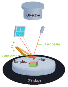
The AFM consists in a microscale cantilever with a sharp tip at its end that is used to scan the surface sample. When the tip gets close to the sample surface, forces between the tip and the sample lead to a deflection of the cantilever. Typically, the deflection is measured using a laser spot reflected from the top surface of the cantilever into a photodiode.
AFM advantages in biology :
- High resolution imaging
- Imaging topographic surfaces at nanoscale (mechanical properties of a sample surface)
- Mechanical characterization of cells or tissue with elastic modulus
An elastic modulus, in Pascal (Pa), is a quantity that measures an object or substance’s resistance to being deformed elastically (i.e., non-permanently) when a stress is applied to it. A stiffer material will have a higher elastic modulus.
Sample surface’s measurements
Topography and mechanics
BioMeca has developed an innovative approach to extract topographic data and localize the area of interest with high-resolutive images. We obtain these images with the Contact Mode.
Another AFM mode, named QNM (Quantitative Nanomechanical Mapping) allows us to associate topography and forces data.
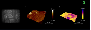
A : human skin optic image B: same skin topographic image obtaines with AFM contact mode
C : topographic image including mechanical data obtained with QNM mode
High-resolution 2D imaging and 3D reconstruction
Thanks to its nanoscale resolution, BioMeca’s technology provides a unique solution addressing multiples applications :
- Interaction drugs/polymers
- Membranes morphological response to nanoparticules
- Structural properties of membranes before/after drugs treatments
- Antibodies morphology
- Etc.
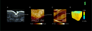
A : skin section optical image B : Contact mode image (topography + adhesion) C : high resolutive imaging of collagen network in dermis D : reconstruction via mechanical approach developed by BioMeca (BioMeca Analysis v3.1®)
3D cartography and multi layers analyses
Combining 3D images obtained with confocal microscopy and data from elastic modulus allows us to study multi-layers tissues.
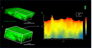
A : skin explant confocal images A’ : 3D extraction of a zone of interest. B : mean elastic modulus of the area
Extraction of metric data and 3D reconstruction
Mecametrix® developed by BioMeca allows to get a 3D modeling organ with data from confocal microscopy and from the biological sample volume or contact surface data.
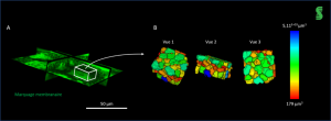
A : skin explant’s representation acquired with confocal microscope B : zone of interest extraction and 3D reconstruction