
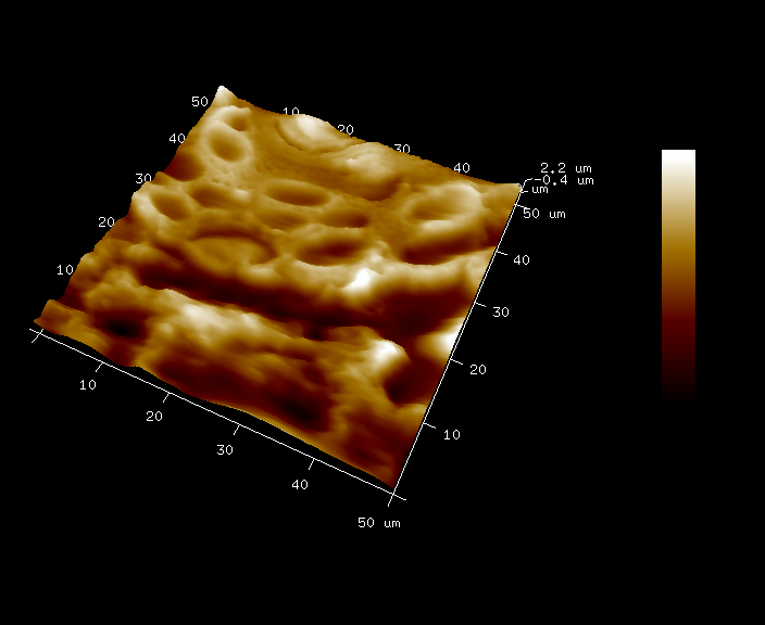
Desquamation
May 2022
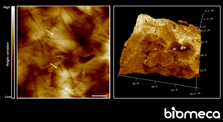
Skin explant topography performed by high-resolution AFM imaging. Some corneocytes involved in the desquamation process are visible (arrows).
Scale bar: 20µm.
Regeneration
April 2022
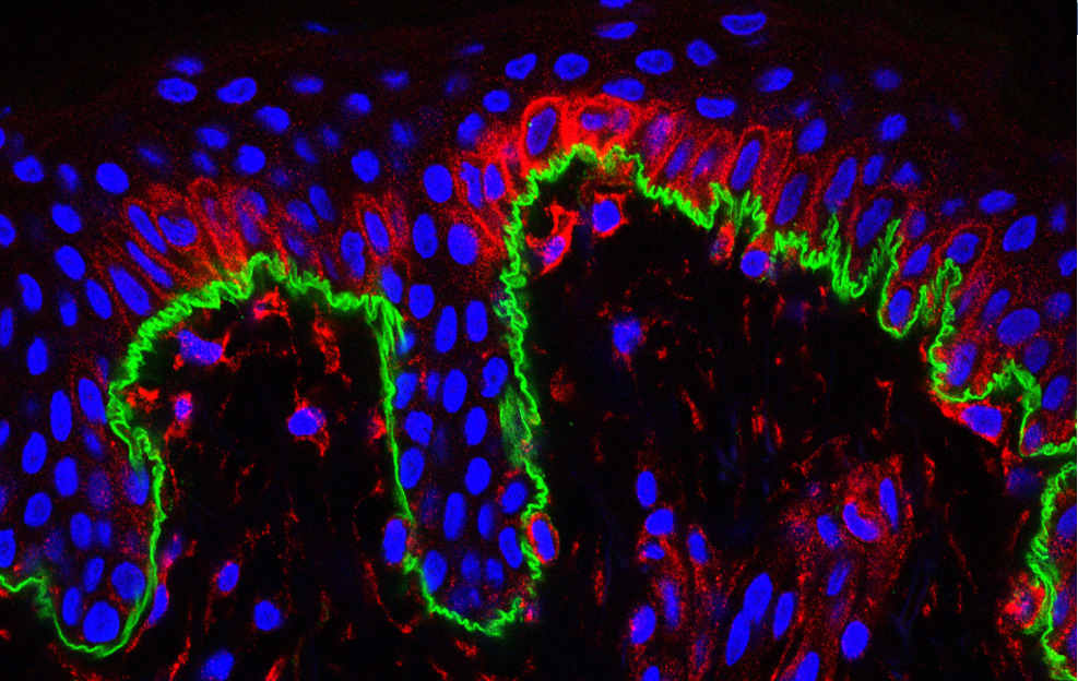
Coupling Atomic Force Microscopy and confocal microscopy to showcase skin regeneration
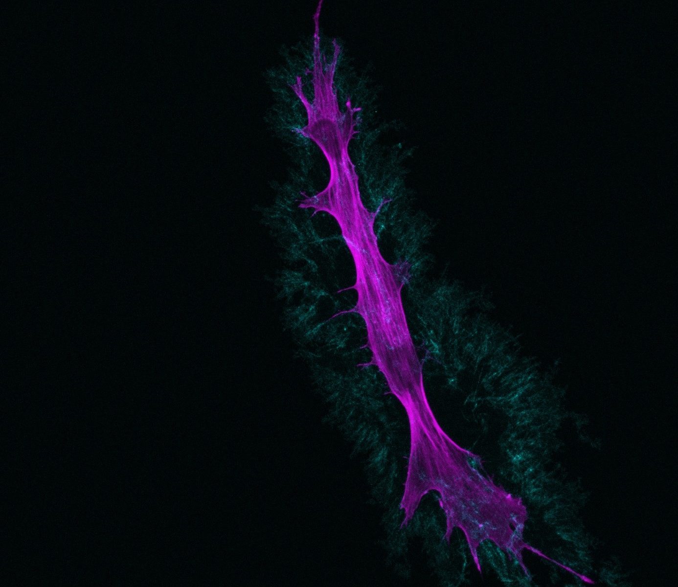
Fibroblasts
September 2021
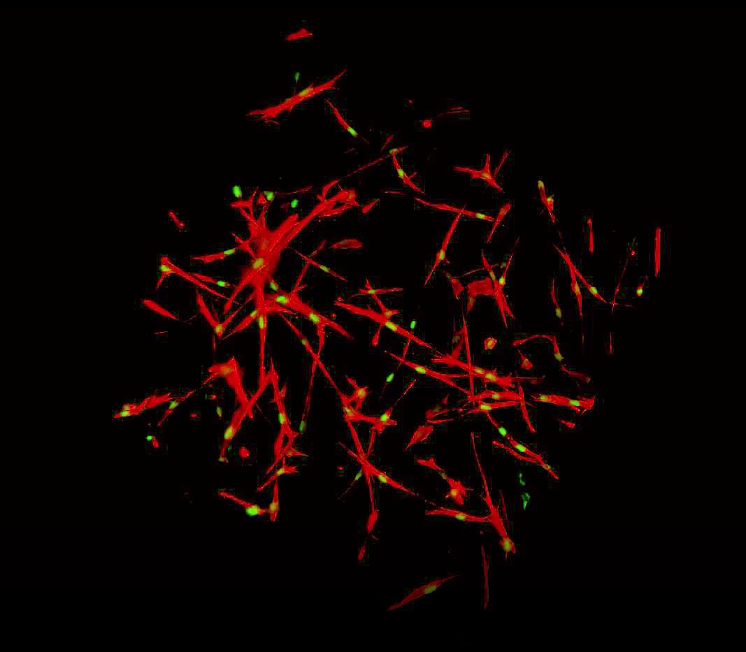
Culture of fibroblasts in collagen discs, allowing the culture of fibroblasts in a three dimensional system, alternative to the classical two dimensional culture system.
Nuclear (green) and actin cytoskeleton (red) labeling.
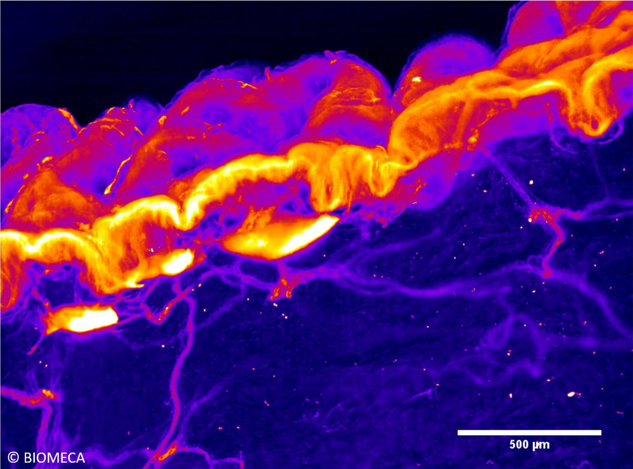
Dermal-epidermal junction
June 2021
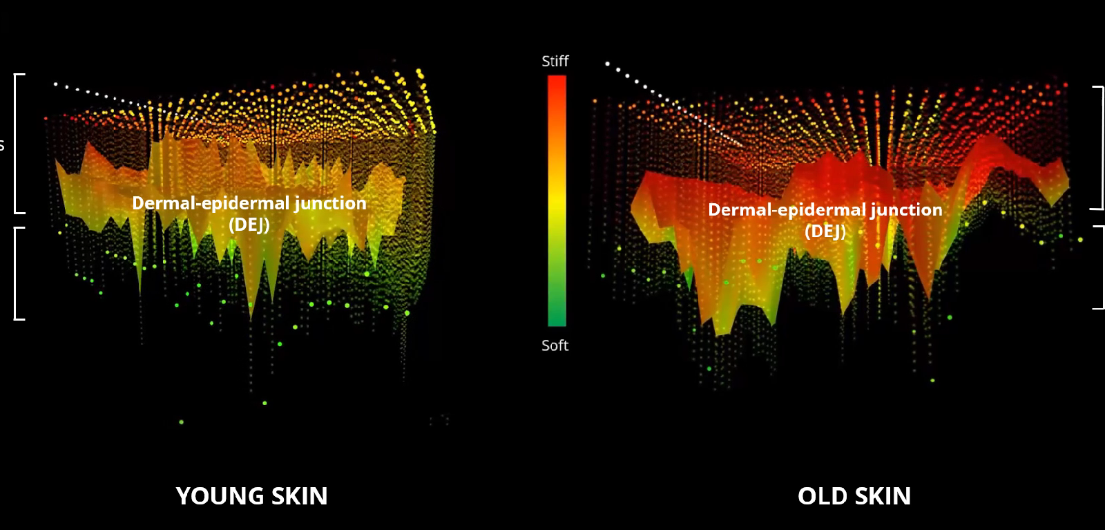
3D force map of dermal-epidermal junction (DEJ) evolution over time with Atomic Force Microscopy (AFM).
Each pixel corresponds to a stiffness value:
🟢 indicates a low stiffness
🔴 indicates a high stiffness
🟠 indicates an intermediate value
A new solution to assess skin ageing focusing on DEJ.
Skin compartments
February 2021
AFM topographic image of skin from abdominoplasty, highlighting the main skin compartments:
👉 At the top of the picture, epidermis with its 3 layers (granulosa, spinous and basal) and at the very top, stratum corneum.
👉 Dermis, at the bottom of the picture, with above epidermis-dermis junction-containing area.
Fibroblasts
November 2020
High-resolution image of a fibroblast using confocal microscopy (for phalloidin labeling of actin) and second harmonic microscopy (for collagen).
Carried out on an uncoated petri dish, with a collagen neo-synthesised by fibroblast.
Skin aging process
July 2020
Microcirculation study through skin capillaries high resolution imaging by light sheet fluorescence microscopy in order to better understand skin aging process.
Discover our solutions

Anti-aging and regenerating effect
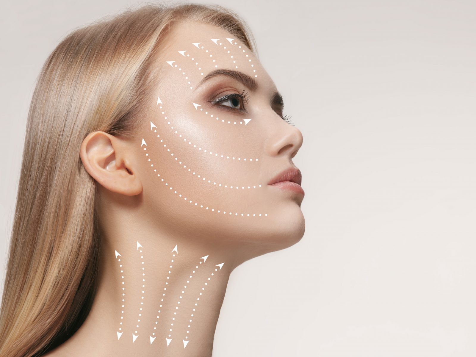
Smoothing effect