Scientific publications
1. Stem integrity in Arabidopsis thaliana requires a load-bearing epidermis
Mariko Asaoka, Mao Ooe, Shizuka Gunji, Pascale Milani, Gaël Runel, Gorou Horiguchi,Olivier Hamant, Shinichiro Sawa, Hirokazu Tsukaya and Ali Ferjani.
2. βig-h3-structured collagen alters macrophage phenotype and function in pancreatic cancer
Sophie Bachy, Zhichong Wu, Pia Gamradt, Kevin Thierry, Pascale Milani, Julien Chlasta, and Ana Hennino.
3. Stiffness measurement is a biomarker of skin aging in vivo
Runel G, Cario M, Lopez-Ramirez N, Malbouyres M, Ruggiero F, Bernard L, Puisieux A, Caramel J, Chlasta J, Masse I.
4. Biomechanical Properties of Cancer Cells
5. Gradient in cytoplasmic pressure in germline cells controls overlying epithelial cell morphogenesis
Lamiré L-A, Milani P, Runel G, Kiss A, Arias L, Vergier B, de Bossoreille S, Das P, Cluet D, Boudaoud A, Grammont M. November 30, 2020.
6. Gene profile of zebrafish fin regeneration offers clues to kinetics, organization and biomechanics of basement membrane.
Nauroy P, Guiraud A, Chlasta J, Malbouyres M, Gillet B, Hughes S, Lambert E, Ruggiero F. Matrix Biology
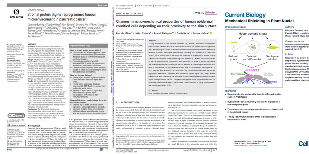
7. Changes in nano-mechanical properties of human epidermal cornified cells depending on their proximity to the skin surface
Milani P, Chlasta J, Abdayem R, Kezic S, Haftek M. J Mol Recognit. 22 mai 2018;e2722.
8. Variations in basement membrane mechanics are linked to epithelial morphogenesis
Chlasta J, Milani P, Runel G, Duteyrat JL, Arias L, Lamiré LA, Boudaoud A, Grammont M. Development 2017 : doi: 10.1242/dev.152652
9. Stromal protein βig-h3 reprogrammes tumour microenvironment in pancreatic cancer
Goehrig D, Nigri J, Samain R, Wu Z, Cappello P, Gabiane G, Zhang X, Zhao Y, Kim IS, Chanal M, Curto R, Hervieu V, de la Fouchardière C, Novelli F, Milani P, Tomasini R, Bousquet C, Bertolino P, Hennino A.
10. Mechanical Shielding in Plant Nuclei
Goswami R, Asnacios A, Milani P, Graindorge S, Houlné G, Mutterer J, Hamant O, Chabouté M-E.
11. Changes in nano-mechanical properties of human epidermal cornified cells in children with atopic dermatitis
12. KATANIN-dependent mechanical properties of the stigmatic cell wall mediate the pollen tube path in Arabidospis
13. Action of Ultra-low Dose Medicine on Oxidative Stress and Cell Stiffness of Microglial Cells In Vitro with Actin Filaments Reorganization
GAËL RUNEL, ANNE PAUMIER, JUSTINE VERRE, ANNA CATTE, SANDRA TRIBOLO, JULIEN CHLASTA, NAOUAL BOUJEDAINI
14. EASYSTIFF®, a portable and innovative device able to separately analyze each skin compartment for the evaluation of mechanical properties
Posters
Visible facial pores : new insights for their assessment and tightening treatment
EXSYMOL
Valenti L., Bliaux J.-P., Lomonte E., Morand B., Guglielmi J., Coste E., Prouheze P., Markioli P.-G.
Today, many people use close-up photos or videos in social media. However, these tend to reveal facial imperfections such as visible pores, complexion, wrinkles and fine lines. In the skin, there are two types of pores: sweat pores, and “visible” pores, where sebum secretion occurs. These small orifices are part of the pilosebaceous apparatus. Intrinsic (genetic predisposition, aging, hormones, hyperseborrhea…) and extrinsic factors (UV, xenobiotics…) are described as being able to cause dilation or enlargement of the facial pores, making them visible to the naked eye [1]. This aesthetic imperfection generates in some people a phobia qualified as “porexia” by dermatologists. The trend for selfies and the quest for an “Instagram Face” exacerbates this feeling. Partly for these reasons, treating visible pores has become a concern for both cosmetics and dermatology. Furthermore, the exact causes of appearance of visible pores are still widely debated in the literature[1,2]. […] In this study, we present an original approach for assessing the effect of a new silanol SiA (combination of adenosine and a core of organic silicium (MTS)) in the dermis and on the specific structures around the pore. We then present the resulting effects of a topical treatment with this silanol on skin biomechanical properties, on skin relief and on pore perception.
Study of the dermal-epidermal junction organization and mechanical properties by Atomic Force Microscopy
EXSYMOL
Nicolaÿ J-F, Valenti L, Markioli P-G, Runel G, Chlasta J. P-S4062
The dermal-epidermal junction (DEJ) is a complex and highly organized structure, which primary function is to anchor the epidermis to the dermis. The DEJ thus preserves skin integrity and participates to its mechanical properties. It also has an important metabolic role as it controls exchanges between epidermis and dermis (water, nutrients, growth factors,…). Atomic Force Microscopy (AFM) is an innovative technique that enables to determine DEJ molecular organization and mechanical properties (mechano organization al analysis).How some animals regenerate missing body parts is not well understood. Taking advantage of the zebrafish caudal fin model, we performed a global unbiased time-course transcriptomic analysis of fin regeneration. Biostatistics analyses identified extracellular matrix (ECM) as the most enriched gene sets. Basement membranes (BMs) are specialized ECM structures that provide tissues with structural cohesion and serve as a major extracellular signaling platform. While the embryonic formation of BM has been extensively investigated, its regeneration in adults remains poorly studied. We therefore focused on BM gene expression kinetics and showed that it recapitulates many aspects of development. As such, the re-expression of the embryonic col14a1a gene indicated that col14a1a is part of the regeneration-specific program. We showed that laminins and col14a1a genes display similar kinetics and that the corresponding proteins are spatially and temporally controlled during regeneration. Analysis of our CRISPR/Cas9-mediated col14a1a knockout fish showed that collagen XIV-A contributes to timely deposition of laminins. As changes in ECM organization can affect tissue mechanical properties, we analyzed the biomechanics of col14a1a-/- regenerative BM using atomic force microscopy (AFM). Our data revealed a thinner BM accompanied by a substantial increase of the stiffness when compared to controls. Further AFM 3D-reconstructions showed that BM is organized as a checkerboard made of alternation of soft and rigid regions that is compromised in mutants leading to a more compact structure. We conclude that collagen XIV-A transiently acts as a molecular spacer responsible for BM structure and biomechanics possibly by helping laminins integration within regenerative BM.
Exploration of biological and mechanical characteristics of a reconstructed dermal model
IFSCC (18-21 SEPTEMBER 2018)
Mariette V, Fernandez E, Runel G, Chlasta J, Marull-Tufeu S, Laperdrix C. P-S4048.
The 1970s saw the advent of cell culture methods, and the development of reconstructed dermal models, with the Bell’s model. Nowadays, full reconstructed skin models are highly developed. They reflect a large part of the biomechanical properties of the skin that are mainly explained by the contractile power of fibroblasts (Bell et al, 1979). We interested in Bell’s model with fibroblasts conditioned by Transforming Growth Factor (TGFb1, 5 ng/ml). TGFb1 is known to stimulate cellular function and to improve contractile power of fibroblasts as it is implicated in healing process. Resistance and elasticity seem also to be improved but nothing was measured and described in literature. We decided to characterize mechanical properties of Bell’s model studying its behavior towards compression and decompression forces, representative of resistance and elasticity. Technics like indentation allowed us to measure the model hardness. Then, these physical results were conforted with microscopic measurements and observations using AFM technic. It highlighted cohesion forces between fibroblasts and the surrounding matrix, inside the dermal model. Finally, the study was completed by fibroblasts distribution observation within the matrix by two-photon microscopy.
Medaka fish as a good model to study skin aging by Atomic Force Microscopy
CENTRE DE RECHERCHE EN CANCÉROLOGIE DE LYON (CRCL)
Runel G, Lopez N, Cario-André M, Chlasta J, Masse I.
Studying skin aging in animal models, either to better understand age-related physiological changes or to find novel molecules that could act on aging, remains relatively long and costly. Fish could be appropriated models for this aim since skin structure is sufficiently similar to mammals one. Indeed, fish has been shown remarkably useful to investigate pigment biology (Schartl and Larue 2016). In particular, the Japanese Medaka, a small fish model with attractive experimental characteristics, could be of interest for studying aging since it accumulates over lifespan some biomarkers currently associated with aging features in others vertebrates. However, little is know about aging of the Medaka fish skin especially in its biomechanical properties and skin architecture. Our work aimed at studying Medaka skin aging at molecular, cellular and biomechanical levels.
Highlight the efficacy of your cosmetic products with Atomic Force Microscopy
BIOMECA
Drillat A, Paillier C, Runel C, Chlasta J
Atomic Force Microscopy (AFM) is composed by a sharp tip attached on a flexible cantilever which is used to indent or to scan sample surface. When a known force is applied, the cantilever bends; this process is monitored with the use of a laser beam reflecting from the top of the cantilever into a photodetector. From the process mentioned above, we obtain force–displacement curves which give us accurate results about mechanical properties of the sample. It also provides probe displacement on the surface to generate high resolution imaging. By using high resolution surface imaging, traction force assay and Stiffness Tomography (ST), BioMeca has developing a non-invasive method to measure the mechanical properties of biological samples (skin, hair etc.. ): hydration, elasticity, ageing, UV radiation effects, blue light, etc..
Structural and biomechanical properties of a novel 3D microdermis model: the spheroid
GATTEFOSSÉ
Lorion C, Lopez-Gaydon A, Bonnet S, Drillat A, Milani P, Bechetoille N
Spheroids as microtissues are a powerful alternative to standard 2D cell culture for in vitro studies. 3D scaffold-free spheroids are formed within a few days from a cell suspension using hanging drop technology. The advantages of spheroids exclusively composed of fibroblasts rely on the physiological production of the extracellular matrix thanks to the aggregative capability of fibroblasts to self-assemble in a round tridimensional structure. The microdermis presents a complex tissue organization that closely mimics the architecture and composition of the human dermis in vivo. The aim of this study was to characterize structural and biomechanical properties of spheroids composed of normal human fibroblasts at 8 and 15 days of culture.
Communications
Application of Atomic Force Microscopy to skin biology (only available in french)
Drillat A, Runel G, Chlasta J, Milani P
La peau constitue l’organe le plus étendu du corps humain. Elle joue un rôle de barrière, mais sert également à communiquer avec l’environnement extérieur. Elle est caractérisée par ses capacités d’élasticité et ses propriétés mécaniques qui lui sont propres et lui confèrent ses fonctions. La peau humaine est composée de plusieurs couches, chacune avec une structure et une fonction unique. Comprendre le comportement mécanique de ces différentes couches est important pour la recherche clinique et cosmétique, à titre d’exemple, pour le développement de produits de soins et la compréhension des affections cutanées.
Corneocytes nano-mechanical properties changes indicate alteration of the skin barrier


