
Atomic Force Microscopy (AFM) allows studies of biological samples and molecules with atomic resolution under near-physiological condition.
Cells, under external stimuli or any physiological modifications, bear physicochemical properties changes that directly affect physiological processes. Among them, cell morphology, elasticity and adhesion properties are crucial processes in carcinogenesis.
That’s why AFM is a relevant tool for studying mechanical properties of cells for high resolution research and especially in cancer research.
Firmness and shape of cells fit their surrounding environment to ease metastasis. Even adhesion between cells is reduced to promote invasion of cancer cells. AFM meets its whole potential to study the morphology of these cells and measure their mechanical properties at nano-scale resolution.

Main AFM advantages
- 3D imaging at nano-scale structures
- Under-near physiological conditions (no damage, no staining)
- Records in real time
- Structural and mechanical information at the same time
- Functionalization of the AFM probe
This last advantage is great to identify specific molecules or interaction forces such as ligand-receptor interactions.
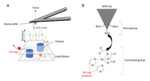
A : Mapping adhesion forces on membrane proteins using FD-based AFM. When recording an AFM topograph an approach (blue) and retraction (yellow) cycle between AFM tip and biological sample is performed for every pixel of the image. In each cycle, the cantilever deflection and the distance travelled by the AFM tip is monitored and transformed into an approach and retraction Force Distance (FD) curve
B : Functionalization of AFM tip (tris-Ni2+-NTA)
Zoom on which differences AFM can reveal in cancer cells
Cells morphology
Some studies have showed differences between cells from leukemia patients. AFM revealed needle-like structures on the surface of leukocytes and a higher roughness of cell surface of white blood cells. A classical optical microscope can’t detect this kind of morphological characteristics (cf. “A mechanical biomarker of cell state in medicine” Di Carlo (2012)).
AFM is also able to analyze membrane surface and highlight cell membrane differences between normal cells and cancer cells.
Mechanical properties
We know that homeostasis of tissues, migration, division and cell growth are processes for which mechanical strength of cells are involved as well as cancerous cells stiffness is not the same than normal stiffness or even metastatic cancer cells. Thus, in measuring stiffness, AFM can help to :
- Discriminate cells,
- Better understand tumour cells microenvironment involved in tumour escape.
Combined with a confocal laser microscope, we’ll also be able to directly observe morphological and mechanical cell changes of cancer development.
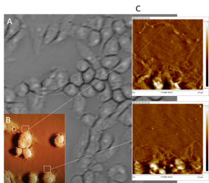
A : optical image of cells
B : AFM topographical image of cells
C : topographical image of the matrix synthetised by the cells obtained by AFM
AFM key assets in cancer research
- Diagnosis for single cells cancer in observing changes and differences between cancerous and non-cancerous cells
- Developing anti-cancer drugs in investigating spreading and interaction processes between cells.
AFM is an exciting technology in both cancer research, diagnosis and clinical treatment. With topographical mechanical measurement, molecular recognition, primary cell detection, and tissue characterization, AFM represents a great multifuctionnal tool for studying physical properties at nano-scale and opens a new door for nanomedecine in oncology.
Discover our study cases

Tissue stiffness

Tumor escape and extracellular matrix

Skin disorders
Follow our other news

The relationship between skin elasticity and viscoelasticity
 24 January 2024
Are you a smoker? Here’s how it affects your skin!
29 November 2022
Why are simplicity and multifunctionality new trends in the beauty market?
19 October 2022