Understanding the mechanical and adhesion properties of skin surface
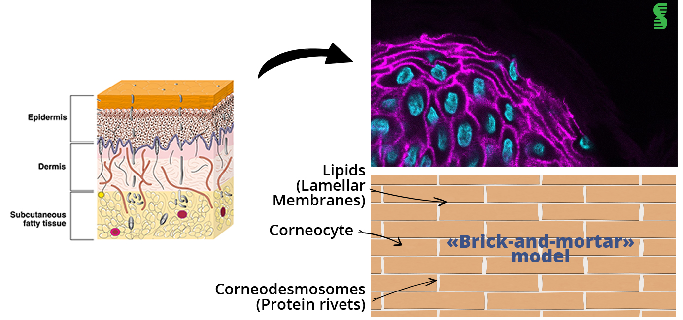
The protective function of biological surfaces that are exposed to the exterior of living organisms is the result of a complex arrangement and interaction of cellular components. This is the case of the most external layer of skin, the stratum corneum (SC). It is composed of corneocytes, elementary ‘flat bricks’ embedded in a lipid matrix. These corneocytes are arranged in layers and adhere to each other through junctions called corneodesmosomes.

Stratum Corneum lipids also play a key role in saving the water barrier of the skin.
The composition of the SC lipid matrix is dominated by three lipid classes: cholesterol, free fatty acids and ceramides. It has been proved that ceramides level decreases with increased age (1) as well as in skin health conditions as atopic dermatitis (2).
Adhesion and mechanical interaction between lipid layers are essential characteristics to highlight the protective function of the Stratum Corneum but are generally poorly understood. New insights provided by Atomic Force Microscopy (AFM) pave the way for new cosmetic products development.
Measuring adhesion forces
Adhesion forces are involved in a mechanism by which cells interact and bind to neighbouring cells, substrates and matrix. It is a necessary mechanism for structural integrity and barrier function of the epidermis.
Some studies have explained the role of individual cell-cell junctions and their protein complexes in regulating epidermal development and function. Adhesion is provided by specialised cellular connections, mainly adhesion junctions (AJ), desmosomes, and tight junctions but not only. Contribution of the skin surface film is also essential. We will focus here on the outermost layer, the Stratum Corneum, specialized to protect from water loss, dehydration, and toxin entry into the body.

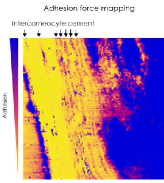
Adhesion force mapping of stratum corneum generated from AFM
Why Atomic Force Microscopy (AFM)?
Many factors influence the adhesion measured with AFM, including not only chemical composition, surface morphology, but also water absorption.
Adhesion is so directly correlated to the composition of skin surface lipids. More the skin will contain intercellular lipids, more it will be adhesive as they act like a water-retaining.

Roughly, the system relies on the displacement of the tip toward the sample and monitors the deformation of the sample induced by the force applied on the cantilever (nano-indentation). Then, the system records both the approaching and retracting curve, as force applied versus distance travelled by the piezo. The retracting curve is usually the one that holds information regarding the strength of the interaction; a minimum in the retracting curve is associated to the adhesion.
Adhesion is measured at a nanoscale level by using the pull-off force when the tips withdraws the sample at the skin surface area end suddenly repelled. Then, according to Hooke’s law, adhesion force is characterized by the bending of the cantilever.
Thus, using a simple AFM measurement, it is possible for the skin to measure adhesion forces. These last ones indirectly correspond to a hydration state, provided by the level of lipids present in stratum corneum.
Atomic Force Microscopy is so versatile that we can as well measure stiffness, elasticity but also adhesion. Such a great tool to characterize skin behaviour and assess active ingredients, formulas or finished products dedicated to maintaining, regulate and beautify our skin.
(1) G Imokawa, A Abe, K Jin, Y Higaki, M Kawashima, A Hidano, Decreased level of ceramides in stratum corneum of atopic dermatitis: an etiologic factor in atopic dry skin?
(2) J Rogers, C Harding, A Mayo, J Banks, A Rawlings, Stratum corneum lipids: the effect of ageing and the seasons
Follow our other news
The relationship between skin elasticity and viscoelasticity
 24 January 2024
Are you a smoker? Here’s how it affects your skin!
 29 November 2022
Why are simplicity and multifunctionality new trends in the beauty market?
 19 October 2022


