
Epidermal differentiation triggers to the formation of dead cells, cornenocytes which regulate epidermis permeability.
Evaluation of skin barrier restoration
BioMeca® provides a solution to evaluate a nourishing cream effect through the study of the structural and mechanical properties of stratum corneum. BioMeca also characterizes corneocytes’ aspect (rugosity, stiffness…) before and after treatment with a cream, to prove a smoothing effect.

Download the full version!
Discover our other solutions
Case studies
Smoothing effect
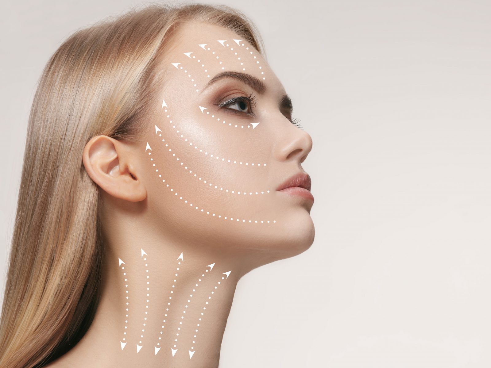
Skin mobilization is the result of the different mobility capacities of the skin and its underlying structures. When the skin is stretched and mobilized, there is an elastic restoring force opposite to the axis of tension,which tends to return the skin to its previous state.
See the study

Case studies
Tensing effect
The dermis is largely composed of dense collagen-rich extracellular matrix (ECM). Dermal collagen represents by far the most abundant ECM protein and constitutes the bulk of skin.
See the study

Case studies
Wound healing
Wound healing is a very complex process, specific to every organism and tissue, and addresses several factors and different cellular and tissular groups.
See the study
Need more innovative cosmetical solutions?