Pancreatic ductal adenocarcinoma (PDAC) is the fourth most frequent cause of cancer-related deaths worldwide and should become the second one hence 2030.
The extracellular matrix (ECM) in the tumor microenvironment modulates the cancer cell phenotype, especially in pancreatic ductal adenocarcinoma (PDAC), a tumor characterized by an intense desmoplastic reaction.
PDAC is characterized by an intense “desmoplastic reaction”, consisting of the abnormal accumulation of ECM components, mostly collagen fibers. During PDAC progression, the pancreatic tumor microenvironment plays a key role as a modulator of cancer cell phenotype, behavior, and chemoresistance.
In this microenvironment, the stroma is characterized by the development of extensive fibrosis, with stromal components outnumbering pancreatic cancer cells.
Most therapeutic treatments has been developed to target cancer cells but not stroma. For PDAC, it should be very interesting to investigate and understand the role of the pancreatic stroma.
Indeed, this massive stroma promotes immunosuppressive response due to partly physical and functionnal modifications of cancer cells surrouding microenvironment. We chose to study mechanical alterations of this microenvironment to understand their impact on cancer cells and immune response. We also studied tumour stiffness to get insights in its role in tumour progression. Indeed, recent studies showed that softe cells are able to form “invasive tips” that eventually are able to break away and spread to other sites, or metastasize.
BioMeca®’s solutions
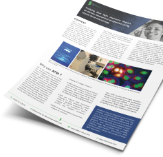
BioMeca® is able to quantify stiffness parameters of pancreatic tumour tissues with AFM (Atomic Force Microscopy). We will carry out experiments on mice pancreatic tumor tissues treated or not with a stromal modulator.
Models : cryosections from treated mice pancreatic tumor tissues
Method : We use AFM mode allowing to obtain high-resolution images and force–volume curves. From them, we build topographic maps and extract mechanical properties of the interest area.
A complete AFM analysis consists in positioning the AFM tip on a specific cryosection area selected with epifluorescent microscope.
Figure 1 : AFM measurement on pancreatic tissue section.
A and B : Fluorescent images of pancreatic tumour section in green and stroma in red.
C : acquisition force-volume curves.
CONCLUSION
Quantitative mechanical data and tumour and stroma areas are then precessed in boxplots graphics to show the stiffness differences between conditions.
References :
Contact us
46 Allée d’Italie
69007 Lyon
France
Interested in this study? Send us a message!
